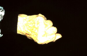
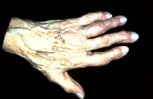
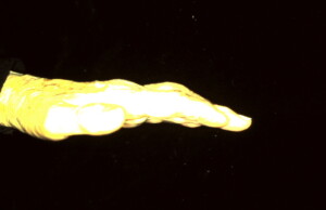
Arthroplasty Proximal Inter-phalangeal Joint Ring Finger
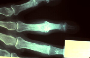 A-P X-ray demonstrating destructive changes of the proximal inter-phalangeal joint of the ring finger. Boney sclerosis and significant bone spur formation evident on the head of the proximal phalanx and base of the middle phalanx.
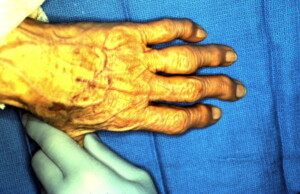 Intra-operative view with osteoarthritic changes of many joints. The ring finger was the most severe and causing the greatest amount of discomfort and functional disability.
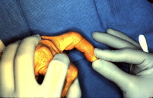 Very limited passive flexion of the Ring finger PIP joint.
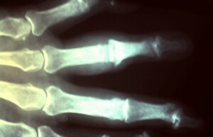 PA view of silicone joint in place.
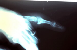 Lateral view of joint in place.
Post operative PA view flexion.
Post operative PA view extension.
Post operative lateral view extension.
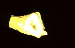 Post operative lateral view flexion.


