Blatt Capsulodesis for S-L Dissociation
Scapho-lunate ligament injuries are quite difficult to treat and results are variable. Acute tears of the scapho-lunate ligament should be treated early with either pinning and immobilization or direct repair. Unfortunately these injuries are typically found late and can not be repaired primarily. Typically the torn ligament results in dissociation or separation of the scaphoid and lunate bones and the scaphoid rotates towards the palm called rotatory subluxation of the scaphoid. the capitate another wrist bone drives itself between the scaphoid and lunate further separating the scaphoid and lunate. This derangement of the wrist bones causes mal-alignment and therefore irregular wear and tear on the cartilage surfaces and ultimately arthritis.
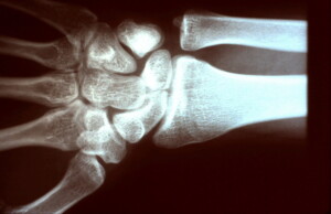 PA radiograph showing signet ring sign of scaphoid because it is flexed or rotated down towards the palm.
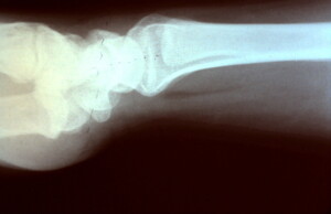 Lateral radiograph demonstrating increased scapho-lunate angle secondary to the rotatory subluxation of the scaphoid.
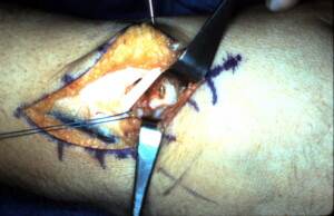 Dorsal intra-operative view of right wrist with extensor pollicis longs being retracted, a prolene suture within a proximally based capsular flap which will be anchored to the hole which was made and is visible at the distal end of the scaphoid. A K-wire is also seen which is holding the scaphoid in anatomic position correcting the rotatory subluxation.
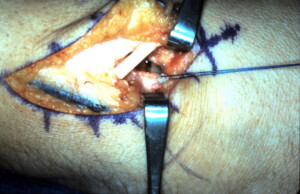 Closer view of the capsular flap being pulled distal.
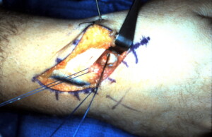 Two free Keith needles are placed through the drill hole in the distal pole of the scaphoid and brought out volarly so the prolene suture within the capsular flap can be anchored to the scaphoid.
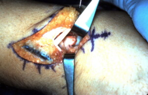 The flap is anchored to the distal pole of the scaphoid and once healed will prevent rotatory subluxation of the scaphoid.
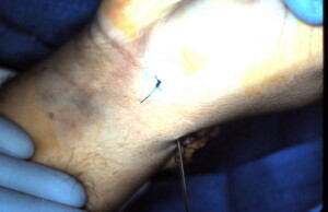 The prolene suture is seen tied over xeroform gauze and a polypropylene button on the palm side of the wrist at the scaphoid tubercle.
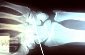 Post op radiograph with K-wire in place maintaining scaphoid position until capsular flap has healed to the bone.
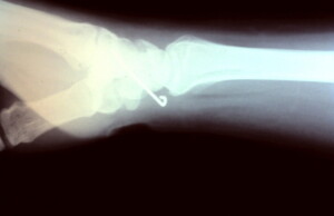 Lateral radiograph showing correction of the scapho-lunate angle.


