

Scaphoid Fracture Non-Union – Repair with Bone Grafting
Fractures of the scaphoid are relatively frequent wrist injuries. They are frquently overlooked because the area may not be noticeably swollen or intensely painful and if non-displaced may not be immediately evident on plain radiographs. It is frequently misdiagnosed as a wrist sprain and remain undiagnosed for months or years, leading to long-term consequences of painful arthritis. That is why a diagnosis of a wrist sprain should have repeat radiographs performed up to three weeks following the injury to pick up this overlooked condition to prevent the adverse long term sequelae.
A scaphoid fracture is usually caused by a fall on the outstretched hand or the result of a similar traumatic force, and occurs most often in young, active patients pursuing athletic sporting activities.
The scaphoid bone has a precarious blood supply and fractures of the waist may disturb the native blood supply to the proximal pole and result in avascular necrosis of that segment of bone, Therefore, even with a proper, timely, diagnosis and treatment it can lead to a failure of the fractured bone to heal resulting in a nonunion.
Diagnosis is made with a thorough physical examination and possibly additional imaging studies such as a bone scan, computed tomography (CT), or magnetic resonance imaging (MRI).
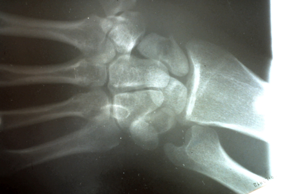
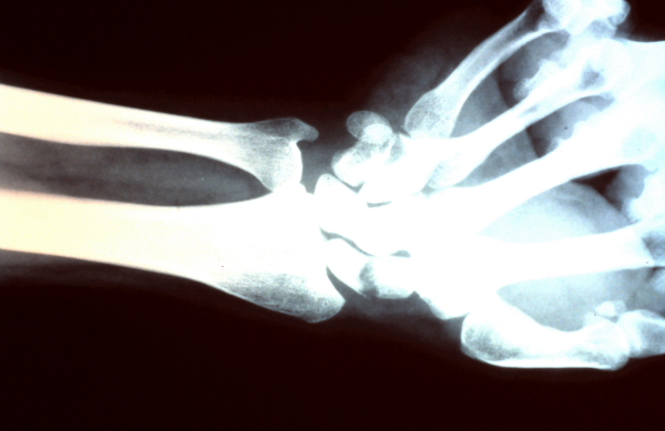
PA X-ray showing transverse waist non-united fracture of the scaphoid which failed to heal with an extended period of immobilization.
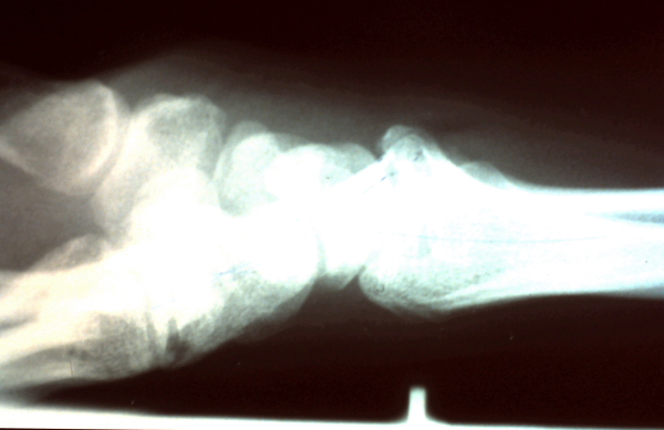 Lateral x-ray of same fracture.
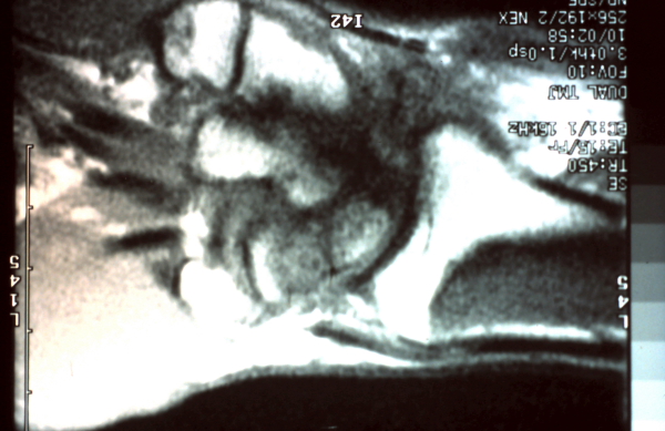 MRI was performed to determine if the proximal pole was viable and amenable to bone grafting.
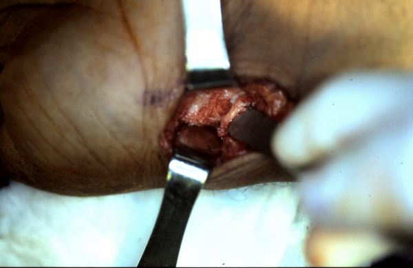
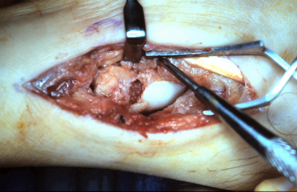
 The tourniquet is released to confirm bleeding from the proximal pole of the scaphoid to ensure healing following bone grafting.
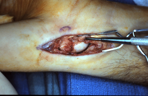
 After the fracture site is debrided of fibrous tissue the defect is measured.
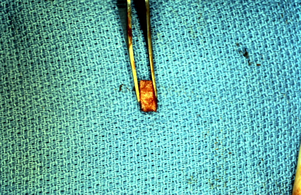 A appropriate sized cortico-cancellous bone auto-graft is harvested to be inserted into the defect.
 The defect is packed with cancellous bone and the cortico-cancellous piece will be placed preventing volar collapse and a humpback deformity of the scaphoid.
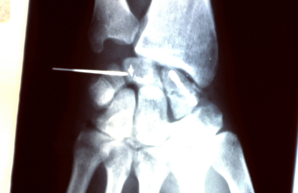 The fracture site was immobilized with a Herbert screw.
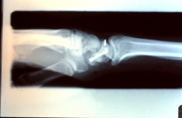 Lateral view with healed fracture evident and humpback deformity corrected.
Case #2 Non-Union Scaphoid Fracture
This patient’s diagnosis was missed and presented in a delayed fashion and underwent bone grafting and internal fixation to achieve a solid bony union.
 Scaphoid non-union evident on plain PA radiograph.
Bone graft inserted into defect.