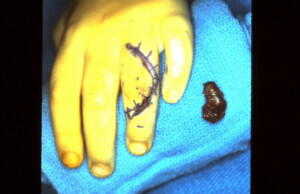
Hemangioma
Subcutaneous hemangiomas are the fourth most common tumor of the hand. They consist primarily of a benign proliferation of blood vessels within the soft tissue. The palm is the most common location. Progressive enlargement of the lesion and throbbing pain are the most common symptoms. General characteristics of a hemangioma include, readily compressible, poorly defined, bluish discoloration, subcutaneous mass that distend when venous return is obstructed and contract when the extremity is elevated. Hemangiomas of the hand are generally surgically excised, with the tributory vessels being identified and ligated as far away from the tumor as possible in order to decrease the chances of recurrence.
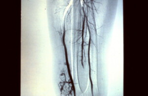 An angiogram demonstrating the anatomy of a hemangioma at the distal medial arm.
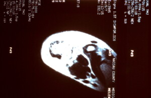 MRI with contrast is performed to further identify the extent of the tumor and isolate its major feeding vessels. Which is sometimes performed in lieu of an angiogram since it is less invasive.
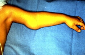 Pre-operative view showing the vascular tumor on the medial aspect of the distal arm.
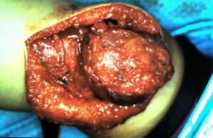 The Hemangioma is nearly completely removed at this stage of the operation. It is imperative to ligate all feeding vessels to decrease the recurrence rate.
Hemangiona of the Index Finger
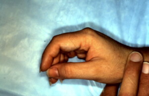 Lateral view of the blood vessel tumor on the volar surface of the index finger.
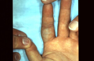 Volar surface showing the typical bluish discoloration.
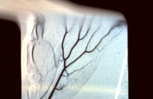 Arterial phase of an angiogram which does not show the vascular lesion filling.
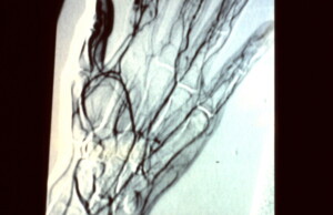 Venous phase of the angiogram demonstrating the lesion on the index finger indicating this tumor has more of a venous malformation character.
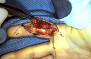 The tumor has been dissected off of the surrounding structures and care being taken to identify the feeding vessels protecting and preserving the digital neuro-vascular bundle.
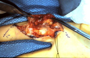 Tumor attached by only small tributaries, which will be ligated and the vascular tumor removed entirely.
Hemangiona of the Ring Finger
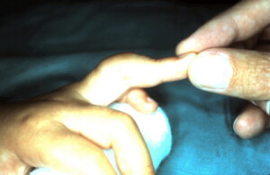 Dorsal aspect of ring finger with vascular tumor evident.


