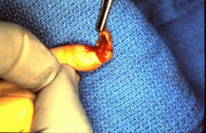
Fingertip Crushing Injury with Nailbed Involvement
Crushed fingertips generally follow a recognizable pattern of injury. Resulting in an avulsion of of the nail plate, laceration to the sterile nail-bed matrix, with or without a fracture of the tuft of the distal phalanx and remain attached by the volar pulp so there is some circulation remaining and have a better chance of survival following re-attachment than would a composite graft if the fingertip was completely amputated.
 Right middle finger crushing injury in a pediatric patient demonstrating avulsion of the nail plate. There is a laceration of the sterile nail bed matrix which is still attached to the undersurface of the nail plate and the distal hyponychium and volar pulp.
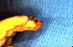 Lateral view showing the laceration to the lateral hyponychium extending towards the volar pulp. This is usually seen on both sides of the digit.
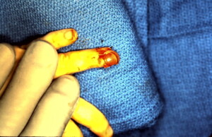 Similar injury to the left ring finger in a child. similarly the nail plate is avulsed and there is laceration of the nail bed matrix with extension on the lateral aspects of the digit. Still attached by the volar pulp.
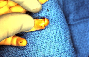 This shows the small remaining skin bridge on the volar surface of the finger which has underlying intact soft tissue attachment maintaining viability.
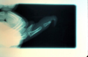 Lateral view of an X-ray reveals a distal tuft fracture which is not uncommon with this type of crushing injury.
The nail plate has been removed and the wound margins debrided. This demonstrates volar soft tissue attachment.
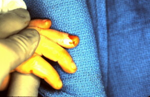 The nailbed and soft tissue has been repaired.
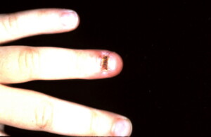 Early post-operative view demonstrating viability of the finger tip. Dry eschar present on the nail bed. The nail will continue to grow and push the scab of the end of the finger.
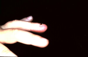 Lateral view. It will take four to six months for the nail to grow back in completely. It is unlikely to have any nail growth deformity unless the injury involves the germinal nail bed matrix.


