Giant Cell Tumor
Giant cell tumors (GCT) are a group of generally benign intra-articular and soft tissue tumors with common histologic features. They can be roughly divided into localized and diffuse types. Localized types include giant cell tumors of tendon sheath and localized pigmented villonodular synovitis, whereas diffuse types encompass conventional pigmented villonodular synovitis and diffuse-type giant cell tumor. Localized tumors are generally indolent, whereas diffuse tumors are locally aggressive. The localized type of the tumor is most commonly found in fingers while subtypes of the diffuse-type GCT may be distinguished as intra-articular and extra-articular. The lesion may appear anywhere in the synovium, but in 80% to 90% of cases, it occurs in the hand joints or tendon synovium, and infrequently in the knee and foot joints. Due to the high recurrence rate of up to 50%, a correct classification of the tumor is essential. As a result of this, and also of the possible malignant degeneration of the tumor, some call the GCT a semimalignant tumor. Complete excision is the preferred treatment for these tumors.
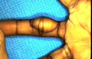 Soft tissue tumor visible on the volar radial surface of the middle finger proximal phalanx.
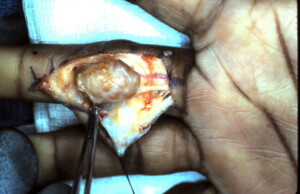 Skin flap elevated exposing the localized variant of a giant cell tumor. Dissection procedes to remove the tumor completely.
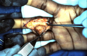 The radial digital nerve which was intimately adherent to the tumor has been freed completely.
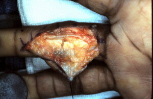 The tumor was found to originate from the synovium of the tendon sheath ad the tendon sheath partially excised and synovectomy performed.
Another Patient with a Localized Giant Cell Tumor
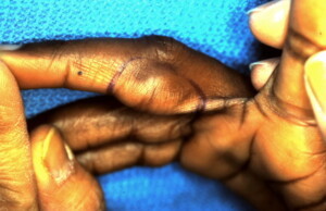
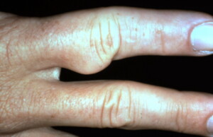 Localized Giant Cell Tumor on the ulnar side of the right index finger.
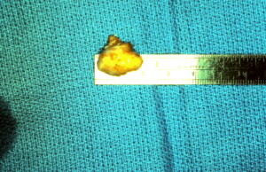
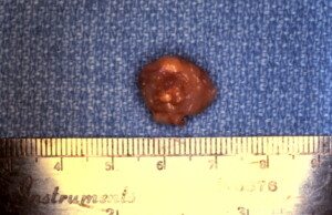 Specimen. This Tumor also originated from the tendon sheath was completely excised with no evidence of recurrence.


